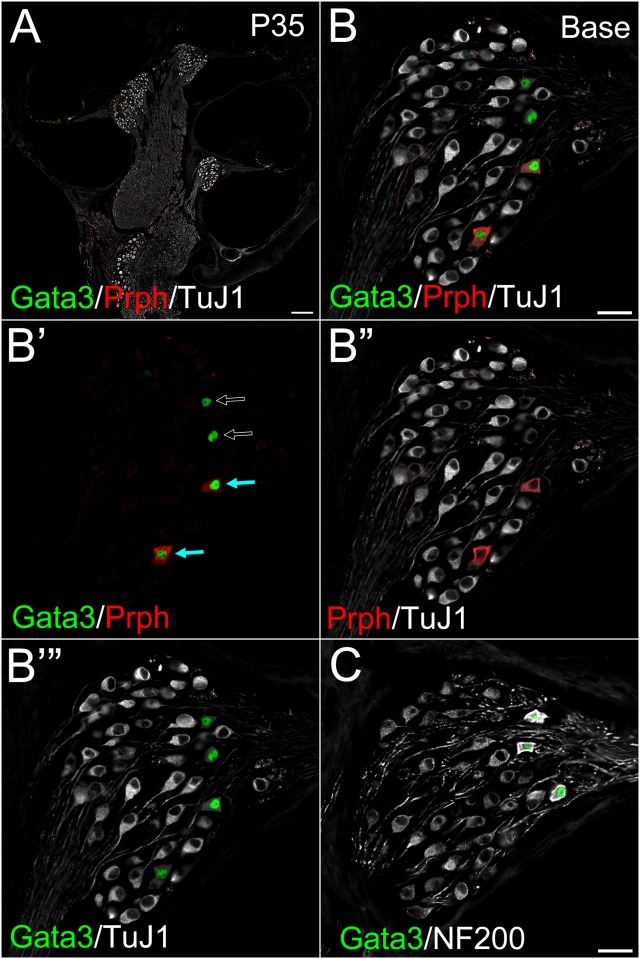
Fig 4. Localization of Peripherin and Gata3 at P35 in the spiral ganglion.
A cochlear cross section of postnatal SG (WT) at P35 immunostained against Gata3 (green), Peripherin (red) and TuJ1 (white) (A−B’”), or Gata3 (green) and NF200 (white) (C). Some Gata3 expressing cells were Peripherin positive, which weakly expressed TuJ1 (indicated by blue arrows in B’). Some Gata3 expressing cells were negative for Peripherin, which weakly expressed TuJ1 (indicated by open arrows in B’). (A−B’”). A: Low magnification view of a cochlear cross section from WT P35 mouse. Gata3 (green) was localized in the nuclei of a few SG cells. Gata3 was also positive in the cells of the organ of Corti including hair cells and supporting cells. B: High magnification view of A, focused on basal SG. B’: The same image as in A, except for TuJ1. B”: the same image as in A, except for Gata3. B’”: The same image as in A, except for Peripherin. C: Another cochlear cross section from P35 mouse. Gata3 (green) strongly positive cells also strongly expressed NF200 (white). Scale bar in A, 100 μm. Scale bars in B and C, 20 μm.

