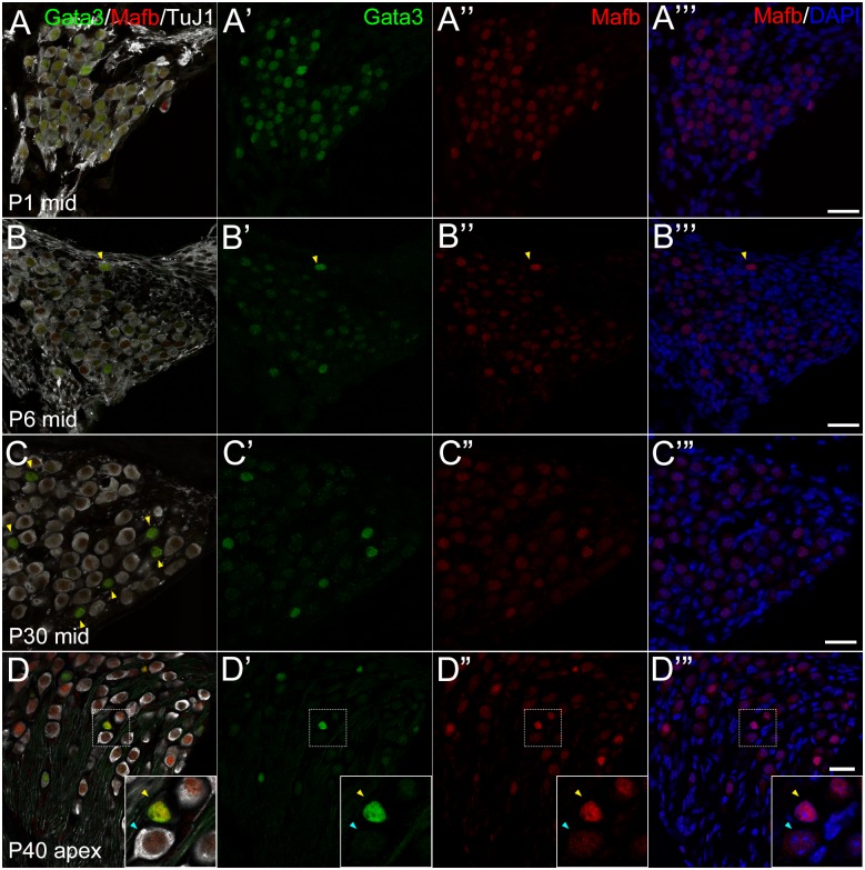
Fig 5. Localization of Mafb and Gata3 at postnatal stages.
Cochlear cross sections of postnatal SG at P1 (A), P6 (B), P30 (C) and P40 (D) immunostained against TuJ1 (white), Gata3 (green) and Mafb (red). Each row shows different stages. A−A”‘: At P1, Mafb (red) was co-localized in the nuclei with Gata3 (green) in TuJ1 (white)-positive PANs. B−B”‘: At P6, like P1, Mafb was localized in most of PANs, and strongly detected in Gata3 strongly positive cells (indicated by yellow arrowheads). C−C”‘: At P30, Mafb was co-localized with Gata3 expression like P6. TuJ1 was negative or weakly positive in Gata3 and Mafb double positive cells indicated by yellow arrowheads in C. D−D”‘: At P40, Mafb expression was the same as P30. High magnification view is shown in the inset, indicating there existed two types of Mafb-positive cells: Gata3 and Mafb double positive cells, which have rather small cell nucleus and did not express TuJ1 (yellow arrowhead); and Gata3 negative, Mafb cytoplasmic positive, and TuJ1 positive cells, which have larger cell body (blue arrowhead). Nuclei were stained with DAPI (blue). Scale bars, 20 μm.

