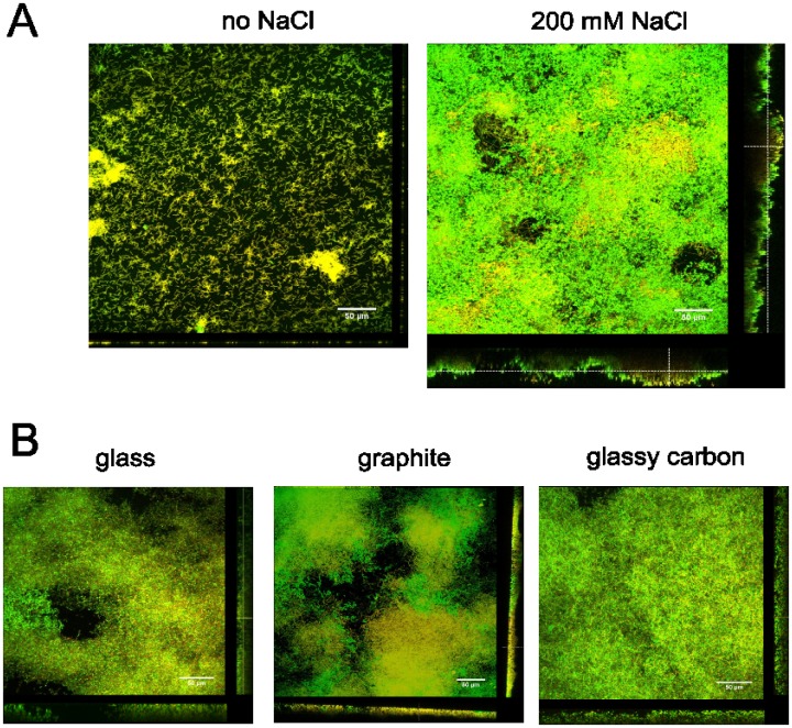
Fig 4. Confocal laser scanning microscopy images of C. ljungdahlii biofilms.
A) Cells were grown in chamber slides without (left) or with (right) the addition of 200 mM NaCl to the medium. B) Cells were grown in tubes, in which a piece of glass (left), graphite (middle) or glassy carbon (right) was placed vertically and to which 200 mM NaCl was added. After 2 days of incubation, the biofilms were stained with live/dead staining as described in the text. The scale bars are 50 μm.

