
Fig. 5.
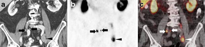
a Coronal CECT; b coronal [18F]DCFPyL PET; and c coronal [18F]DCFPyL fused PET/CT images of a patient with multiple lymph node lesions. There is a large (2.0 cm short axis) lymph node near the region of the bifurcation of the left common iliac artery (arrowheads) that demonstrates intense [18F]DCFPyL PET uptake. Additionally, small (4 and 6 mm short axis) lymph nodes in the right common iliac chain (arrows) are also intensely [18F]DCFPyL-avid.