
Fig. 6.
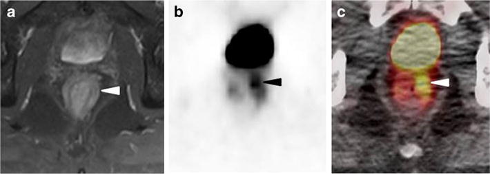
a Axial T2 MRI with endo-rectal probe; b axial [18F]DCFPyL PET; and c axial [18F]DCFPyL fused PET/CT images of the rectal and peri-rectal tissues in a patient with suspected metastatic prostate cancer. Asymmetric radiotracer uptake in the left anterior soft tissues within or adjacent to the rectal wall can be seen (arrowheads) without a corresponding finding on the MRI. This was found to be a site of metastatic prostate cancer on biopsy.