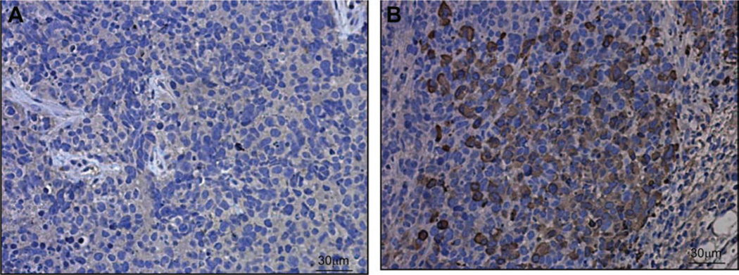
Fig. 6.
Immunohistochemical analysis of H446 human small-cell lung tumor xenografts from mice that were systemically treated with saline or 109 virus particles (vp) per kg Seneca Valley Virus-001 (SVV-001). Mice were killed 24 hours after injection, and tumor sections were subjected to immunohistochemistry using mouse polyclonal antibody against SVV-001 capsid proteins. Cells that are infected by SVV-001 are indicated by brown intracellular staining. A) Image from a tumor of a mouse injected with saline. B) Image from a tumor of a mouse injected with 109 vp per kg SVV-001. One representative image of three experiments is shown.

