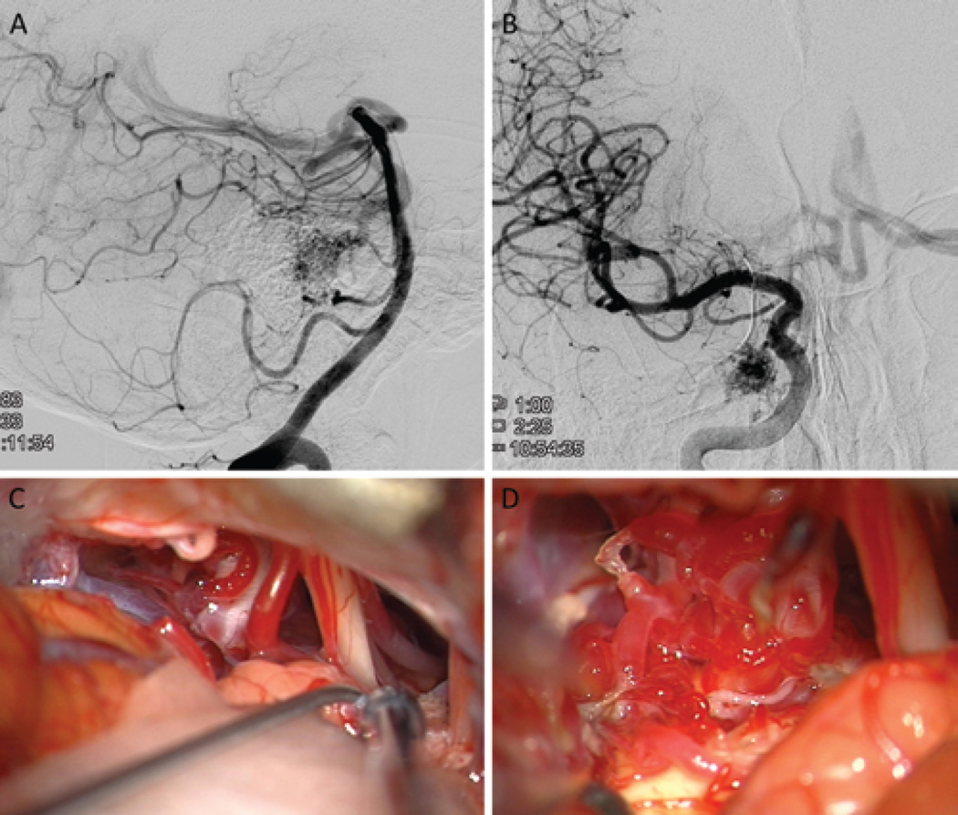
FIG. 3.
A 75-year-old man presented with a remote hemorrhage from a right lateral pontine AVM (supplemented Spetzler-Martin Grade 6 [S1V1E1/A3B0C0; < 3 cm in diameter, deep venous drainage, and in eloquent location/age > 40 years, bled, and compact nidus]), supplied by the AICA (right VA angiogram, lateral view; A) and dural arteries from the meningohypophyseal trunk (right ICA angiogram, anteroposterior view; B). The AVM was approached through a right extended retrosigmoid craniotomy, which exposed CN V, CN VII–VIII (at tips of Rhoton 6 and sucker, respectively), and CN IX (right of sucker tip) (C). The AVM infiltrated the trigeminal nerve root but was mostly lateral to it (D).

