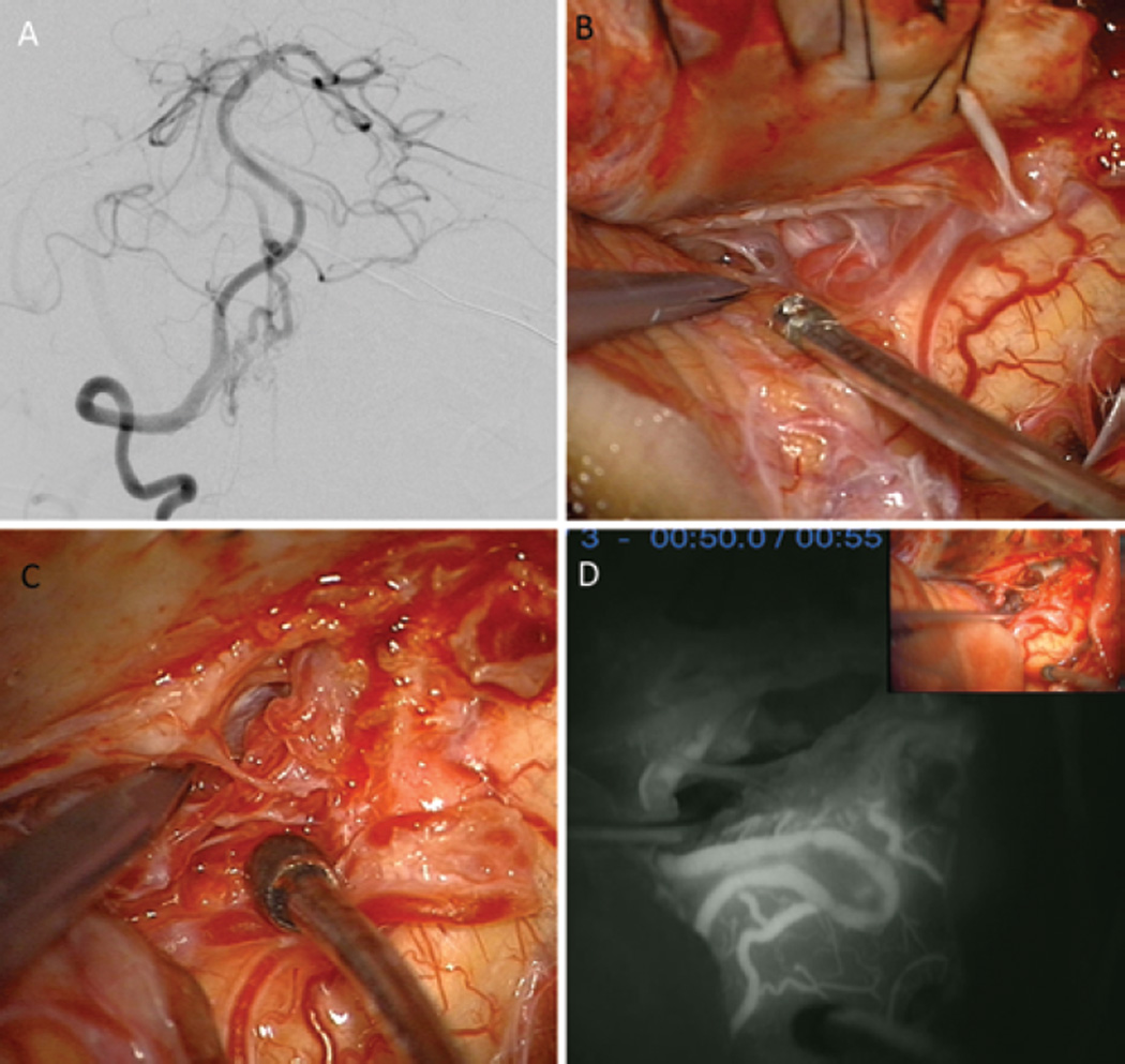
FIG. 5.
A 55-year-old woman presented with an intraventricular hemorrhage from a right lateral medullary AVM (supplemented Spetzler-Martin Grade 6 [S1V1E1/A3B0C0; < 3 cm in diameter, deep venous drainage, and in eloquent location/age > 40 years, bled, and compact nidus]), located on the pial surface of the medulla with dilated draining veins anteriorly. A: The AVM was supplied by PICA branches and the anterior spinal artery (AntSpA) and drained by the MAMedV (right VA angiogram, anteroposterior view). B: A right far-lateral craniotomy exposed the cerebellomedullary fissure and lateral medulla. C: The AVM was on the pial surface of the lateral medulla and extended inferiorly to the cervical spinal cord. D: As PICA branches were interrupted, the PICA separated from the AVM and MAMedV, seen on the anterior medullary surface, darkened. Indocyanine green videoangiography confirmed the absence of arteriovenous shunting. The AVM was occluded in situ, and angiography confirmed complete occlusion.

