. 2017 Jan 16;6:e21510. doi: 10.7554/eLife.21510

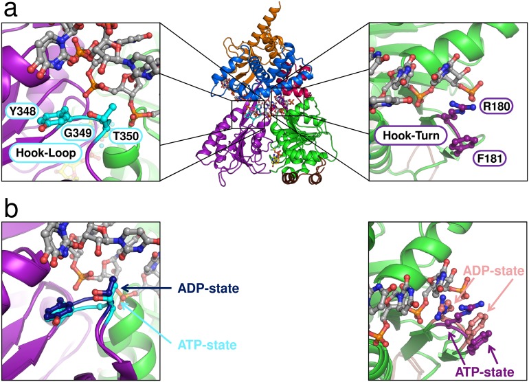
Figure 7. Position of the Hook-Turn and Hook-Loop in ctPrp43.

(a) The localization of the Hook-Turn in the RecA1 domain and of the Hook-Loop in the RecA2 domain in the ctPrp43ΔN•U7•ADP•BeF3- complex structure is shown. Domains are colored according to Figure 1a. (b) Superpositions of the RecA1 and RecA2 domains of the ctPrp43ΔN•U7•ADP•BeF3- and the ctPrp43ΔN•ADP (PDB 5d0u) complexes for the Hook-Turn and Hook-Loop, respectively. The superpositions indicate that the Hook-Loop remains in a highly similar conformation after ATP hydrolysis in contrast to the Hook-Turn which is shifted towards the RNA in the ADP-bound state.

DOI: http://dx.doi.org/10.7554/eLife.21510.022

Figure 7.

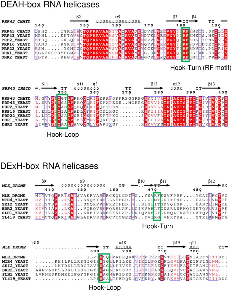
Figure 7—figure supplement 1. Partial sequence alignment of Prp43 from C. thermophilum to all DEAH-box RNA helicases from S. cerevisiae and of MLE from D. melanogaster to all yeast DExH-box RNA helicases.

Figure 7—figure supplement 1.

The amino-acid sequence of ctPrp43 (G0RY84) was aligned to scPrp43 (P53131), scPrp2 (P20095), scPrp16 (P15938), scPrp22 (P24384), scDhr1 (Q04217) and scDhr2 (P36009). The sequence of dmMLE (P24785) was aligned to scMtr4 (P47047), scSki2 (P35207), scBrr2 (P32639), scSlh1 (P53327) and scYL419 (Q06698). The values in parentheses indicate the Uniprot accession numbers (The UniProt Consortium, 2015). The alignment was visualized with ESPript 3.0 (Robert and Gouet, 2014). The Hook-Turn and Hook-Loop motifs are boxed in green.