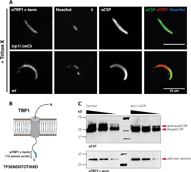
Author response image 2. A peptide antibody designed against the TRP1 N- terminus does not recognize TRP1 by immunofluorescence and western blot.

(A) Immunofluorescence on permeabilized (Triton-X 100) midgut sporozoites of wt and trp1(-)mCh. The staining with αCSP antibodies was included as control to validate the staining procedure. The immunofluorescence signal with the N- terminal αTRP1 was not significantly different from background suggesting non- specific binding. (B) Illustration of TRP1, the location and sequence (15 amino acids) of the peptide used for raising the antibody. (C) Western blot with wt (control) midgut sporozoites. The N-terminal αTRP1 antibody doesn’t recognize any Plasmodium protein. Note that the unspecific binding between 150 and 250 kDa corresponds to a shading of the membrane but not a distinct band that got enhanced during the scanning of the western blot. CSP was used as a loading control (on the right side). Both images correspond to the same sample on the same western blot.
