. 2017 Jan 24;6:e19157. doi: 10.7554/eLife.19157

Figure 4. Complementation of trp1(-) parasites with full-length but not truncated TRP1 restores the wild-type phenotype.

(A) Ratio of hemolymph sporozoites (HLS) to midgut sporozoites (MGS) and (B) of salivary gland sporozoites (SGS) to midgut sporozoites (MGS) for gfp-trp1∆N, gfp-trp1∆C gfp-trp1comp and gfp-trp1 lines in comparison to wild-type (wt) parasites. The bar charts show the mean of four independent countings (10 mosquitoes each) at days 14, 18, 20 and 22 post infection of a selected feeding experiment. For absolute numbers see Table 2. Error bars represent SEM. (C) Sporozoites of gfp-trp1comp in midguts, salivary glands and hemolymph counted over time; 1–2 countings per timepoint. (D) Mechanically ruptured salivary gland releasing gfp-trp1comp sporozoites. Scale bar: 10 µm.

DOI: http://dx.doi.org/10.7554/eLife.19157.012

Figure 4.

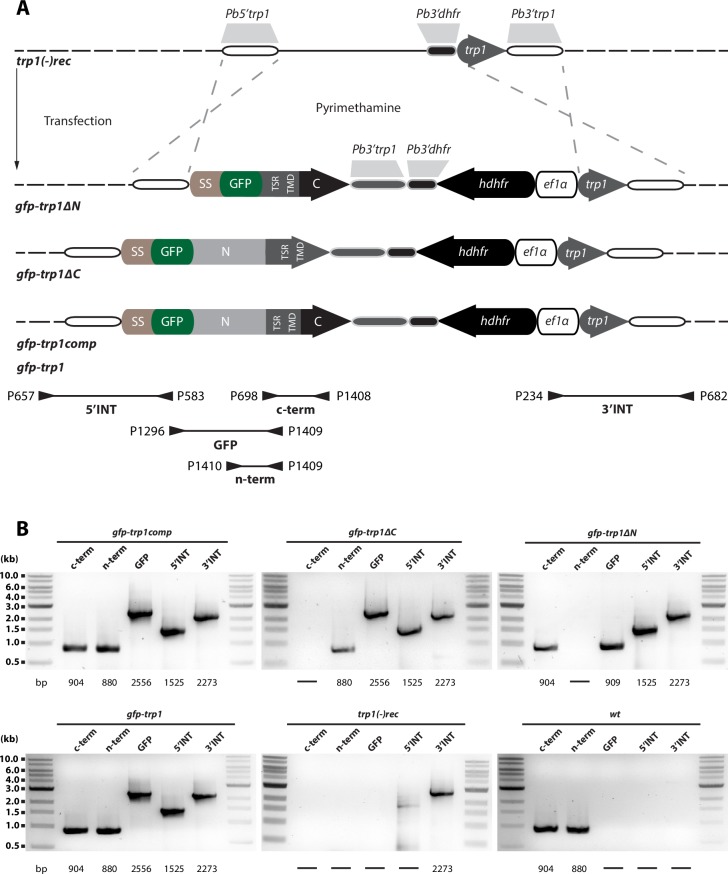
Figure 4—figure supplement 1. Generation and PCR analysis of gfp-trp1comp, gfp-trp1, gfp-trp1∆N and gfp-trp1∆C parasites.

Figure 4—figure supplement 1.

Schematic representation of the complementation strategy with sequences encoding full-length and truncated TRP1 proteins. (A) Complementation was performed with three different constructs (gfp-trp1, gfp-trp1∆N and gfp-trp1∆C). All constructs contained the positive selection marker hdhfr under control of the ef1α promoter and a GFP gene at the N-terminal end, between the sequence encoding the signal peptide and the remaining trp1 ORF. The location of primers and the length of PCR fragments used for genotyping are indicated by arrows and black lines below the scheme. Approximate sizes of the PCR products are indicated below the images. (B) PCR analysis of clonal lines revealed correct integration of the designed constructs. To probe for the absence of deleted sequences, two PCRs specific for the N- and C-terminus of trp1 (n-term and c-term) were performed. The PCR termed GFP amplifies a sequence between GFP and trp1 to test the fusion of both sequences. For comparison, the PCRs for both recipient lines trp1(-)rec and wt are shown.
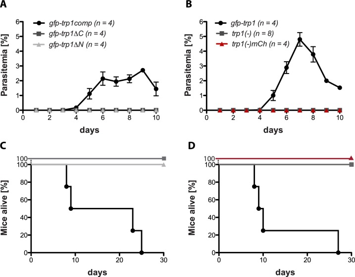
Figure 4—figure supplement 2. TRP1 is essential for transmission by infected mosquitoes.

Figure 4—figure supplement 2.

(A,B) Parasitemia in mice exposed to ten mosquitoes infected (A) with gfp-trp1:compgfp-trp1∆C and gfp-trp1∆N and (B) with trp1(-), trp1(-)mCh and gfp-trp1. Blood stage parasites were monitored for 10 days post infection. The graphs show the mean and the standard error of the mean (SEM) of the parasitemia for all infected mice per group (n). (C,D) The survival of mice infected with gfp-trp1comp, gfp-trp1∆C and gfp-trp1∆N (corresponding to (A)) and with trp1(-), trp1(-)mCh and gfp-trp1 (corresponding to (B)). The viability of all mice was monitored for 30 days post infection.