
Fig. 3. Spatial organization of the Spy-Im7 complex.
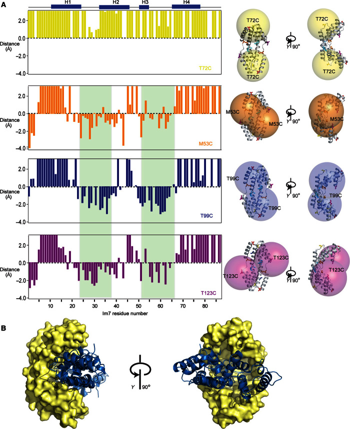
(A) Distance restraints from intermolecular PRE measurements relative to a value of 15 Å. Data of four single mutants of Spy attached with the spin label MTSL are shown: yellow, Spy T72C; orange, Spy M53C; blue, Spy T99C; violet, Spy T123C. Residues 23 to 36 and 50 to 65, which have an averaged distance of less than 15 Å to the spin label center of M53C, M85C, T99C, and T123C mutants, are indicated with green background. Right: For each spin label, spheres with a radius of 20 Å centered around the Cβ atom of each cysteine mutant are shown on the crystal structure of Spy. (B) Rigid-body docking model of the complex of Spy with Im7, based on the PRE data. Three representative ensemble members are shown.