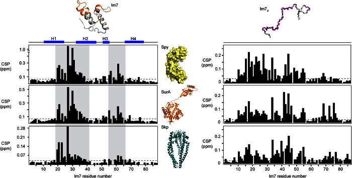
Fig. 5. The interaction sites on Im7 and Im7U for the chaperones Spy, SurA, and Skp.
CSPs of amide moieties plotted against the amino acid residue number of 200 μM Im7 (left column) and 200 μM Im7U (right column) upon interaction with any of the chaperones 240 μM dimeric Spy (top row), 200 μM SurA (central row), or 200 μM trimeric Skp (bottom row). The secondary structure elements H1 to H4 of Im7 are indicated. Segments 19 to 41 and 55 to 66 are highlighted in gray to guide the eye. Structural models are shown for orientation (40, 41). Data in top row are identical to Figs. 2B and 4F.

