
Fig. 3. Localization of nestin+ cells in tendon tissue and endogenous tendon injury repair.
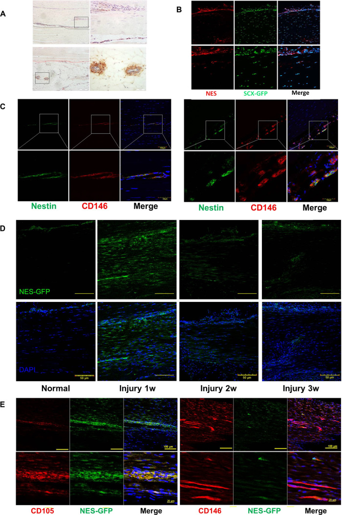
(A) Immunofluorescence staining of nestin expression in the human Achilles tendon endotenon (region between the black dashed lines) and surrounding blood vessels in the peritenon (area below the red dashed line). Scale bars, 200 μm (right); 50 μm (left). (B) nestin expression in Scx-GFP mouse Achilles tendon. (C) Nestin and CD146 expression in human Achilles tendon. Scale bars, 100 μm (top); 50 μm (bottom). (D) Nes-GFP expression in normal and injured Achilles tendon 1, 2, and 3 weeks after surgery (n = 5). Scale bars, 50 μm. (E) Immunofluorescence staining of CD105 and CD146 expression at the injured tendon of Nes-GFP mice 1 week after injury (n = 5). Scale bars, 100 μm (top); 20 μm (bottom).