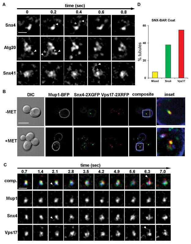
Figure 2. Snx4 family proteins coat endosome-derived membrane tubules.
(A) Micrographs of Snx4-2xGFP, Atg20-2xGFP and Snx41-2xGFP decorated endosomes captured by time-lapse fluorescence deconvolution microscopy. Galleries are representative of SNX-BAR coated endosomes with an associated tubular endosomal network. Arrowheads point to tubules of interest and open arrows (<) indicate apparent fission events. Images were captured at a single focal plane and acquired at 200 msec intervals. (B) Micrographs showing cells expressing Mup1-mTagBFP2, Snx4-2xGFP, Vps17-2xRFP in methionine depleted media. Within 15 minutes of the addition of 20 μg/ml methionine, a subset of Snx4 and Vps17 (a retromer subunit) endosomes accumulated Mup1. Pearson’s correlations of Snx4 and Vps17 is Rave=0.5 (n=20) (C) Micrographs showing distinct tubules emanating from Mup1-mTagBFP2, Snx4-2xGFP, Vps17-2xRFP endosomes. Endosomes were captured by three-color time-lapse fluorescence deconvolution microscopy. An example of a tubule decorated by only Snx4 is shown at 2.1 sec, a tubule decorated by only Vps17-2xRFP is shown at 6.3 secs. For visual contrast, individual colors are shown in gray scale. (D) Quantitation of the different types of SNX-BAR-coated tubules. Of 100 tubules analyzed, 40% were decorated with only Snx4, 57% were decorated with only Vps17, and 3% were decorated with both.

