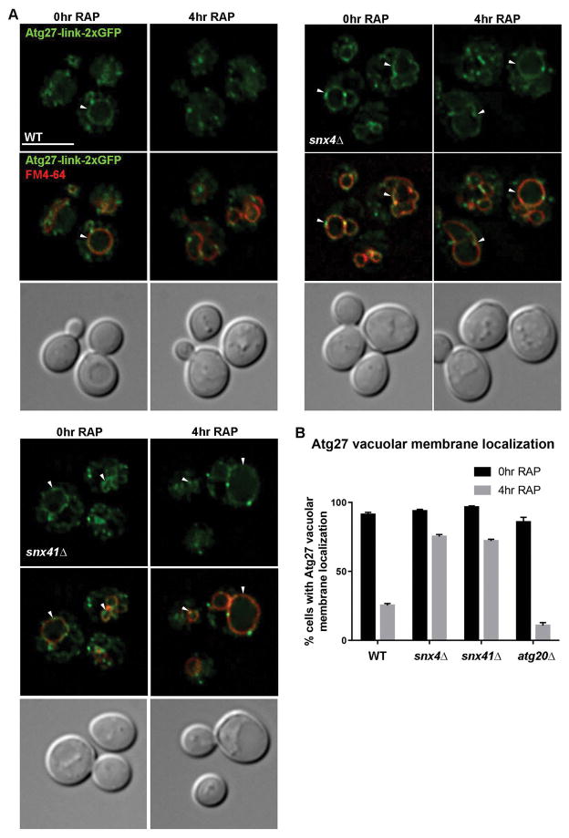
Figure 7. Atg27 fails to mobilize from the vacuole membrane in response to rapamycin treatment in snx4Δ and snx41Δ cells.
(A) Micrographs of cells expressing Atg27-link-2xGFP in wild-type, snx4Δ (B), and snx41Δ (C) cells. Cells were grown to early log phase and incubated with rapamycin (0.2μg/mL) for 4 hr to induce autophagy and labeled with FM4-64 dye as described. Approximate middle slices of deconvolved Z stacks are shown. Vacuoles are identified with arrows. The scale bar indicates 5 μm. (D) Quantification of percent of cells with vacuolar membrane localization of Atg27-link-2xGFP as measured by continuous colocalization of Atg27-link-2xGFP with FM4-64. Error bars indicate SEM of three independent experiments.

