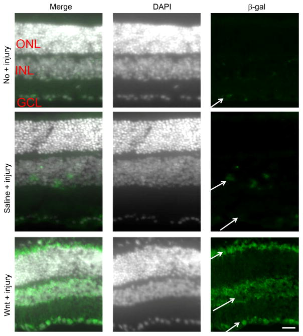
Fig 1. Localization of Wnt signaling.
The Tcf/LacZ transgenic Wnt reporter mice were used to localize cells that are responsive to Wnt3a stimulation. DAPI-stained nuclei are shown in white to demonstrate the retinal layers. Uninjured eyes had barely detectable basal Wnt signaling in the GCL, indicated by low levels of β-gal detection (green; arrows). However, increased expression of β-gal was detected in injured retinas that received saline injection (saline + injury), notably in the INL and GCL, and Wnt3a injection induced prominent Wnt signaling in the INL and GCL, indicated by increased immunostaining of β-gal (Wnt3a + injury). ONL, outer nuclear layer; INL, inner nuclear layer; GCL, ganglion cell layer. Scale bar = 50μm

