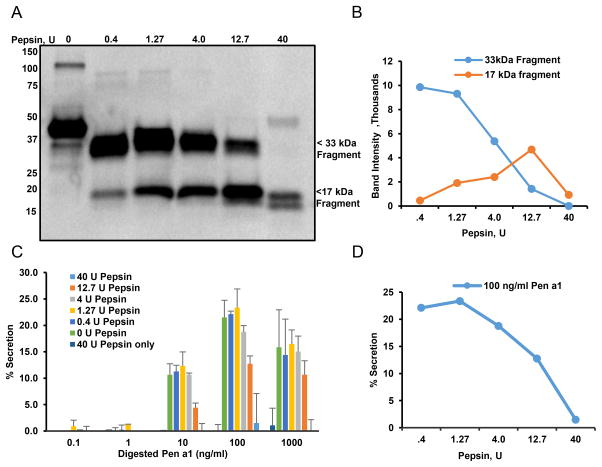
Figure 4. Effect of pepsin digestion on IgE binding to Pen a 1 and basophil degranulation.
A) Immunoblot analysis of IgE from serum of atopic individual binding to rPen a 1 digested with increasing concentrations of pepsin for 10 min at 37oC. Bound IgE was detected using HRP conjugated anti-IgE. B) Quantification of two major fragments obtained with rPen a 1 digestion in SDS-PAGE (B). C) hRBL-2H3 cells were primed with serum of atopic individual and challenged with rPen a 1 digested with increasing concentration of pepsin (concentrations shown in legend). D) Quantification of secretory responses induced by 100 ng/ml rPen a 1 digested with increasing concentrations of pepsin from plot B.

