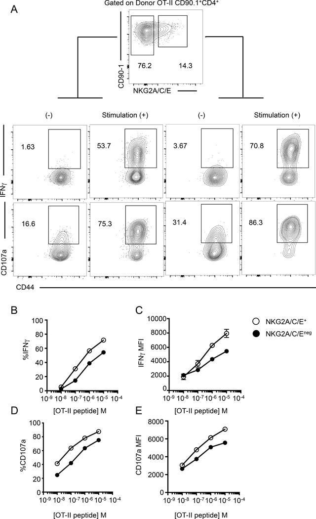
FIGURE 6.

ThCTL show increased IFNγ secretion and degranulation than the non-ThCTL lung CD4 T cells. Lung OT-II.Thy1.1 CD4 T cells were isolated 8 dpi and stimulated with peptide pulsed activated B cells. (A) Representative staining plots of IFNγ and CD107a expression of donor cells after 10−5 M OT-II peptide stimulation. (B) Quantification of the percent IFNγ producing OT-II CD4 T cells gated on NKG2A/C/E+ (open circle) or NKG2A/C/Eneg (closed circle). (C) Median fluorescence values of IFNγ, gated on IFNγ+ cells of indicated populations. (D) Quantification of percent CD107a+ cells. (E) Median fluorescence values of CD107a, gated on CD107a+ cells. (CD4 T cells isolated from pooled lungs, representative of 2 independent experiments, n=5 mice each).
