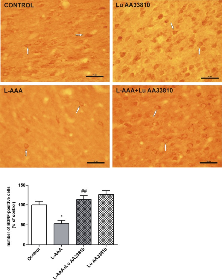
Fig. 5.
(Upper panel) Representative microphotographs of coronal sections of the rat brain showing the expression of BDNF-positive cells in the mPFC of the experimental groups. Many BDNF-positive cells are seen in the control rat. Gliotoxin L-AAA (100 μg/2 μl, twice) induced a strong decrease in the number of BDNF-ir cells, while Lu AA33810 (10 mg/kg, i.p.) reversed this effect. Arrows point to some of BDNF-positive cells. Calibration bars 50 μm. (Bottom panel) A histogram showing the effect of L-AAA and Lu AA33810 administered alone or in combination on the number of BDNF-ir cells in the rat PFC. Data are expressed as % changes vs. control. Values represent the mean ± SEM (n = 5–6 rats per group) and were evaluated by two-way ANOVA, followed by the Bonferroni multiple comparison test. *p < 0.05 vs. control; ## p < 0.001 vs. L-AAA-treated group

