Fig. 2.

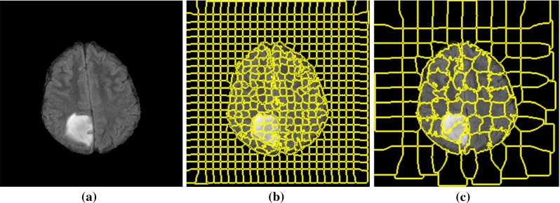
Example of superpixel segmentation with different window sizes: a original MRI FLAIR image with a grade II tumour, b superpixel segmentation with (initial grids ) and , c superpixel segmentation with (initial grids ) and

Example of superpixel segmentation with different window sizes: a original MRI FLAIR image with a grade II tumour, b superpixel segmentation with (initial grids ) and , c superpixel segmentation with (initial grids ) and