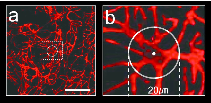
Fig. 1.
Lower (a) and higher (b; dotted square in a) images of confocal laser-scanning microscopy (CLSM) demonstrating the thickness of the myoepithelial cell processes of the rat submandibular gland immunostained for αSMA. For the thickness of a myoepithelial cell process, the length of the portion of the circle 20 μm in diameter with its center at the center of the nucleus intersecting with the process was measured. Bar=50 μm.

