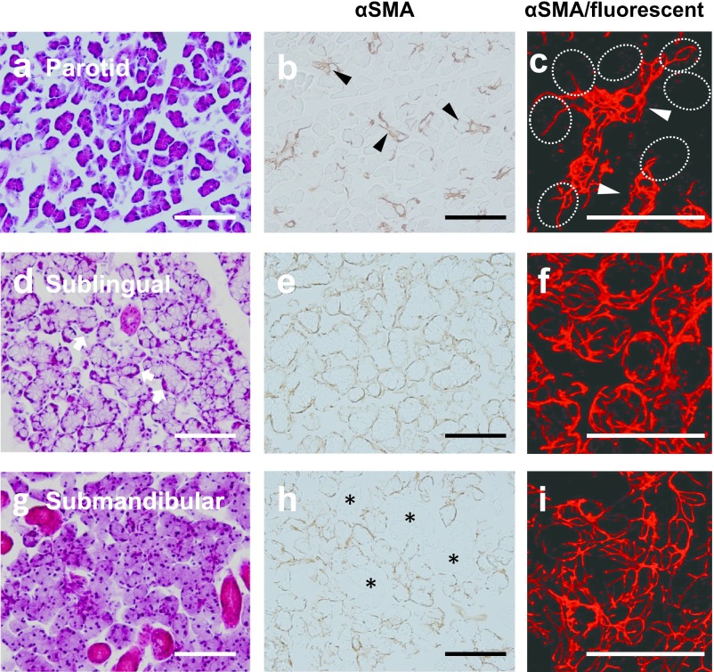
Fig. 2.
Microphotographs of H-E staining (a, d, g), conventional (b, e, h), and higher magnified CLSM immunohistochemistry (c, f, i) stained for αSMA of parotid (a–c), sublingual (d–f), and submandibular (g–i) glands of adult rats. The parotid gland includes developed serous acini, as well as intercalated and striated ducts (a). αSMA-positive myoepithelial cells are rarely on the acini (dotted circles in c), but present along the intercalated duct (arrowheads in b, c). The sublingual gland consists of mixed acini accompanying the serous demilune (arrows in d). Sublingual myoepithelial cells thickly covered mixed acini (e), and their processes are relatively shorter and simpler (f) than those of the submandibular gland (i). The submandibular gland consists of developed serous acini, as well as intercalated, granular, and striated ducts, and lacks mucous cells (g). Submandibular myoepithelial cells cover both acini and intercalated ducts (h), and their processes are relatively longer and more complicated (i) than those of the sublingual gland (f). Granular and striated ducts (*) lack myoepithelial cells. Bar=100 μm.

