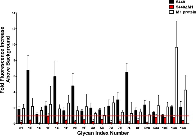
FIG 1 .
The M1 protein is a major lectin of M1T1 GAS clone 5448. Glycan binding of GAS strains 5448 and 5448ΔM1 along with recombinant M1 protein was analyzed using the ProScanArray imaging software ScanArray Express (PerkinElmer, USA), and the data were exported to Microsoft Excel for further analysis. Bacterial and protein binding to a glycan was defined as a value representing a >1-fold increase above the mean background relative fluorescence units (RFU). The mean background was calculated from the average RFU of all empty spots on the array + 3 standard deviations. Statistical analysis of the data was performed by a Student’s t test with a confidence level of 99.99% (P ≤ 0.0001), and only glycans that met these criteria for three biologically independent samples (n = 12 glycan spot replicates) were interpreted as positive binding interactions. Glycan index: 81, Galα1-4GlcNAcβ; 1B, Galβ1-4GlcNAc; 1C, Galβ1-4Gal; 1F, Galb1-3GalNAcb1-4Galb1-4Glc; 1G, Galβ1-3GlcNAcβ1-3Galβ1-4Glc; 1P, Galα1-3Galβ1-4Glc; 2B, Galβ1-6Gal; 2F, Galα1-4Galβ1-4GlcNAc; 4A, GlcNAcβ1-4GlcNAc; 5D, Manα1-3Man; 7A, Fucα1-2Galβ1-3GlcNAcβ1-3Galβ1-4Glc; 7H, Galβ1-4(Fucα1-3)Glc; 7L, Fucα1-2Galβ1-4(Fucα1-3)Glc; 8F, Galβ1-4(Fucα1-3)GlcNAcβ1-6(Fucα1-2Galβ1-3(Fucα1-4)GlcNAcβ1-3)Galβ1-4Glc; 528, Neu5Acα2-3Galβ1-4(Fucα1-3)GlcNAcβ1-3Galβ; 533, GalNAcβ1-4(Neu5Acα2-8)2Neu5Acα2-3Galβ1-4Glc; 10E, Neu5Acα2-3Galβ1-3(Neu5Acα2-6)GalNAc; 13A, ΔUA 2S-GalNAc-4S Na2 (Δ Di-disB); 14A, (GlcAβ1-3GlcNAcβ1-4)n (n = 10).

