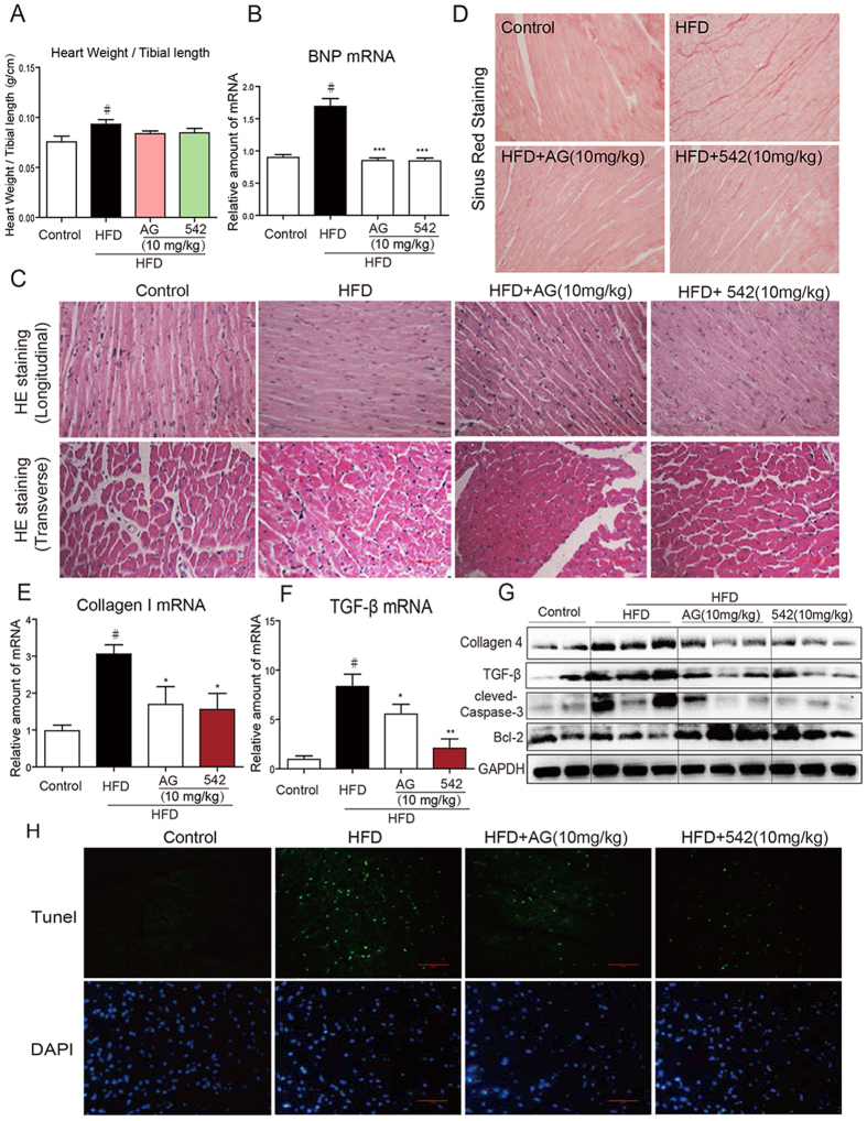
Figure 3. EGFR inhibitors reverse HFD-induced hypertrophic remodeling, fibrosis and apoptosis in C57BL/6 mouse heart.
C57BL/6 WT mice were fed with HFD for 8 weeks, and treated with AG1478 (AG, 10 mg/kg/day) or 542 (10 mg/kg/day) for 8 weeks by oral gavage. (A) Heart Weight/Tibial length ratio (n = 7 per group). (B) qPCR analysis of BNP mRNA levels in the hearts (n = 7 per group). (C) Myocardial histological analysis was performed using H&E staining and representative images were shown (×400). (D) Myocardial fibrosis analysis was detected using Sirius Red staining and representative images were shown. qPCR analysis of TGF-β (E) and collagen1 (F) mRNA levels (n = 7 per group,). (G) The protein levels of collagen 4, TGF-β, cleaved-caspase-3, Bcl-2 were measured by western blot (n = 2 in control group; n = 3 in other groups). The gels were run under the same experimental conditions. Shown are cropped gels/blots (The gels/blots with indicated cropping lines are shown in Supplementary Fig. S4). (H) Representative images of TUNEL/DAPI staining of myocardium were shown. All images were obtained by microscope with original magnification (×400). #p < 0.05 vs. control group; *p < 0.05, **p < 0.01, ***p < 0.001 vs. HFD alone.

