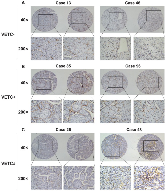
Figure 1.
Vascular pattern types in 168 HCC tissues assessed by immunohistochemical staining for CD34. (A) HCC tissues from two different patients have abundant capillary-like vessels but have absolutely no VETCs, which were classified as VETC-. (B) HCC tissues from two different patients showed distribution of VETC structure in a whole HCC section, which were classified as VETC+. (C) HCC tissues from two different patients displayed mixed vascular structure containing both VETC and capillary-like structure, which were classified as VETC±.

