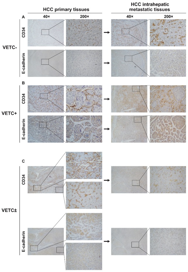
Figure 4.
Vascular pattern type and E-cadherin expression in 50 paired primary and intrahepatic metastatic lesions. (A, B, upper panel) Vascular pattern types of intrahepatic metastatic lesions were similar to their corresponding primary lesions in (A) VETC- and (B) VETC+ group. (C, upper panel) Intrahepatic metastatic lesions from VETC± group showed no VETCs. (A-C, lower panel) In serial sections, E-cadherin expression was highly expressed in VETC positive area, but lowly expressed in VETC negative area in HCC tissues.

