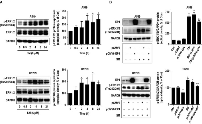
Figure 2.

Solamargine increased the phosphorylation of ERK1/2. (A) H1299 and A549 cells were treated with solamargine (6 μM) in the indicated times, and cell lysate was harvested, and the expression of the phosphorylated and total protein of ERK1/2 was measured by Western blot analysis using corresponding antibodies. GAPDH was used as loading control. (B) A549 and H1299 cells were transfected with control and EP4 expression vectors for 24 hrs before exposing the cells to solamargine for an additional 24 hrs. Afterwards, the EP4 protein, phosphor‐ERK1/2 were determined using Western blot. GAPDH was used as internal control. Values in bar graphs were given as the mean ± SD from three independent experiments. *Significant difference compared with the untreated control group (P < 0.05). **Significant difference from solamargine treated alone (P < 0.05). ERK1/2, extracellular signal‐regulated kinases 1/2.
