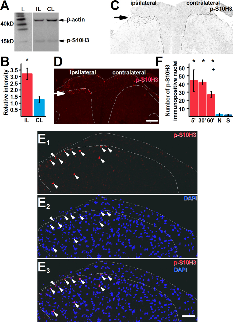
Figure 1. Burn injury induces prolonged up-regulation in p-S10H3 expression in superficial spinal dorsal horn of the spinal cord.
(A) and (B) Burn injury increases p-S10H3 (expected MW: 17kD) expression within 5 minutes in the ipsilateral side (IL) compared to the contralateral side (CL). The expected molecular weight of β-actin is 42 kD. L indicates size marker. *p = 0.006; n = 4. The Western blot images shown in (A) are cropped. (C) and (D) Images of transverse sections of the L4-L5 spinal segments showing both the ipsilateral and contralateral sides. p-S10H3-immunopositive structures in the ipsilateral spinal cord (arrow; scale bar = 200 μm). (E1–E3) Images of the ipsilateral side of a transverse spinal cord section of the L4-L5 segments. Structures exhibiting p-S10H3-immunopositivity in the superficial spinal dorsal horn are invariably positive also to DAPI which binds to adenine-thymine regions of the DNA in the nucleus. Scale bar = 100 μm. (F) p-S10H3 expression lasts for at least 60 minutes (60′) after the injury. 5′, 30′ and 60′, 5, 30 and 60 minutes post-injury, respectively; N, naive; S, 5 minutes after a sham injury. *p < 0.001, + p = 0.001 (n = 4). Dotted and dashed lines indicate the surface of the spinal cord and the white-grey matter border, respectively on each image.

