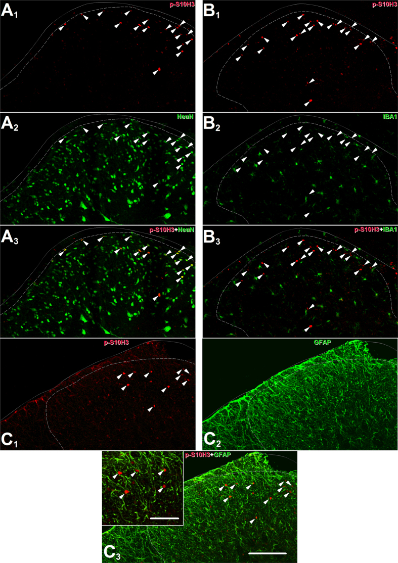
Figure 2. The burn injury-induced up-regulation in p-S10H3 expression occurs in neurons.
(A1–3) Images of the ipsilateral side of a section cut from the L4-L5 spinal segments. p-S10H3-expressing structures (arrows) 5 minutes after burn injury also express the neuronal nucleus marker NeuN. (B1–3) Images of the ipsilateral side of a section cut from the L4-L5 spinal segments. Microglia identified with an anti-ionised calcium-binding adaptor protein 1 (IBA1) antibody do not express p-S10H3 after burn injury. (C1–3) Images of the ipsilateral side of a section cut from the L4-L5 spinal segments. Astrocytes identified with an anti-glial fibrillary acidic protein (GFAP) antibody do not express p-S10H3 after burn injury. (Scale bar, 200 μm; scale bar in inset, 100 μm.) Dotted and dashed lines indicate the surface of the spinal cord and the white-grey matter border on each image.

