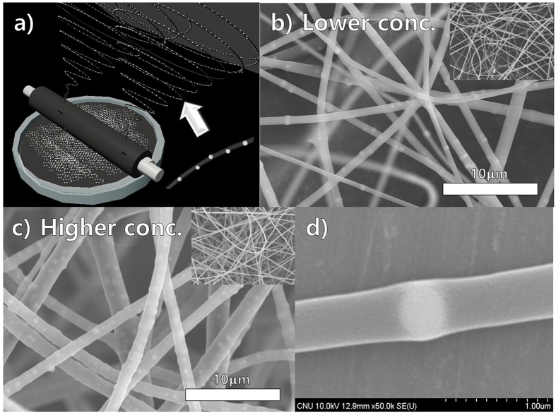
Figure 7.
(a) Schematic diagram of colloidal electrospinning using helically probed rotating cylinder, (b) SEM images represented the colloidal electrospinning using PVP in water and ethyl alcohol with the lower concentration of 500 nm sized silica nano-particles (0.5 g/40 ml), (c) SEM images shows the colloidal electrospinning with high concertation (1.5 g/40 ml), (d) SEM image of single fibers having silica particles with high magnification.

