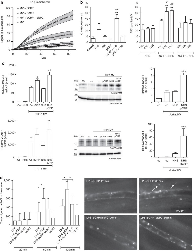
Figure 6. Proinflammatory effects of CRP and microvesicles.
(a) Binding of CMFDA-labelled microvesicles (MV) to immobilized C1q was determined in the presence of pCRP, 1,6-bis-PC-pCRP and mCRP by repeated fluorescence measurements. 1,6-bis-PC is labelled as bisPC. Shaded areas display s.e.m. (n=3). (b) Binding of complement C3 to MV was assessed by flow cytometry with anti-C3b-PE antibodies (n=5), which recognizes C3, C3b and iC3b or three different C3 antibodies that recognize either the C3c part, the iC3b neoepitope or the C3d part, and a second APC-labelled antibody (n=3). pCRP* led to significantly increased C3 deposition that is present on the MV surface mainly in the form of iC3b. No deposition of complement C3 was observed in the absence of NHS or in the presence of heat-inactivated sera (HIS). Displayed are the means and s.e.m. P values were calculated with one-way analysis of variance (ANOVA). **P<0.01 to NHS, ++P<0.01 to HIS, #P<0.05 to mCRP+NHS and ##P<0.01 to mCRP+NHS. (c) Expression of VCAM-1 and ICAM-1 by HUVECs was determined by qRT-PCR (quantitative real-time PCR) and western blot. pCRP+NHS significantly increased the expression of ICAM-1 and VCAM1 on HUVECs in the presence of cell-derived MV. Compared to THP-1 MV (n=4), Jurkat MV led only to a small increase in ICAM-1 and VCAM-1 expression (n=4). Displayed are the means and s.e.m. P values were calculated with one-way ANOVA. **P value <0.01 and ***P value <0.001 compared to NHS. #P value <0.05 compared to THP-1-MV. Uncropped images of western blots are shown in Supplementary Fig. 14. (d) pCRP significantly increases the number of transmigrated leukocytes in LPS-induced inflammation in rat cremasteric postcapillary venules. This effect can be masked by preventing pCRP dissociation with 1,6-bis-PC. Transmigration of leukocytes was analysed by intravital microscopy under superfusion with LPS (25 ng ml−1)±intravenous injection of pCRP (25 μg ml−1) that had been preincubated with 1,6-bis-PC in some groups. After labelling with rhodamine 6G, the number of transmigrated leukocytes was quantified in a visual field of 200 μm in length and of 100 μm in width in the immediate vicinity of a postcapillary venule after 20, 60 and 120 min. Values are mean±s.e.m. of six rats per group; P values were calculated with one-way ANOVA, *P<0.05. Images shown are typical examples for postcapillary venules under LPS and pCRP in the presence and absence of 1,6-bis-PC after 20 and 60 min. Scale bar, 100 μm and applies to the whole panel.

