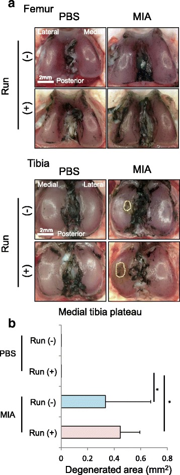
Fig. 3.

Macroscopic observation for articular cartilage. a Macroscopic images of the femoral and tibial articular cartilages stained with India ink. Cartilage erosion is surrounded by a yellow dotted line. b Quantification of degenerated area of the tibial cartilage (n =4, * p < 0.05 by Steel-Dwass test)
