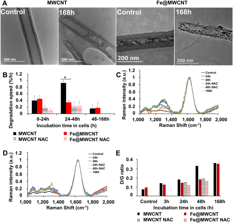
Figure 1. MWCNT and Fe@MWCNT degradation in THP-1 differentiated into macrophages.
Cells were exposed to a 1 μg/cm2 suspension of MWCNTs or Fe@MWCNTs for 24 h. Non- phagocytosed CNTs were removed and (A) TEM observations of extracted MWCNTs or Fe@MWCNTs before (control) and after 168 h aging into cells were performed, showing stimagta of degradation. (B) The degradation speed of MWCNTs and Fe@MWCNTs is calculated for different period of aging in cells in the presence or absence of NAC. Raman spectrum of (C) MWCNTs and (D) Fe@MWCNTs for different times of aging in macrophages in presence or absence of NAC, confirming surface modifications over time. (E) D band/G band ratios of Raman spectra. (*) designates a statistically-significant difference between MWCNT and Fe@MWCNT groups (p < 0.05). ($) designates a statistically-significant difference between MWCNT NAC or Fe@MWCNT NAC group and its respective equivalent MWCNT or Fe@MWCNT group without NAC treatment (p < 0.05). Experiments were repeated at least three times with similar observations in each experiment.

