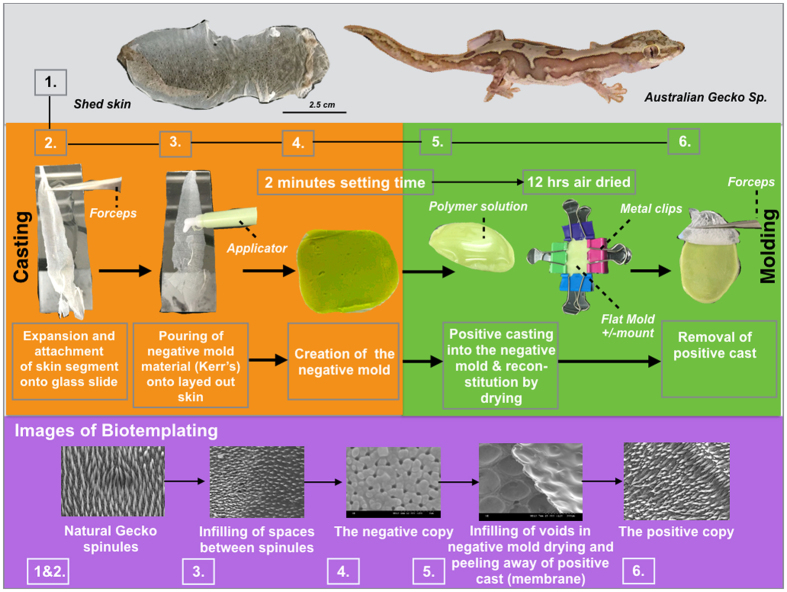
Figure 1. A flow diagram showing the biotemplating methodology for duplicating gecko skin spinules into a small range of biopolymer membranes.
Shed gecko skin was immobilized on a film of water that additionally inflated the spinules into their natural state as seen in the living gecko. Whole shed skin from a single gecko lizard (7–10 cm in length) [step 1] was expanded onto a thin film of water covering a glass microscope slide and dried in place [step 2]. The now flat panel of skin was casted with PVS using a plastic applicator [step 3]. The PVS was hardened within 2 minutes and generated a negative mold [step 4]. The gecko skin was peeled away from the PVS membrane. A polymer solution was added to the gecko skin patterned face of the PVS membrane [step 5]. The polymer solution was air dried for 12 hours and, for replicas made with biomaterial cross-linking agents were added in situ. All membranes were air dried for 1–2 hours prior to use. The positive casted membranes were peeled away from the PVS mold [step 6]. The corresponding Biotemplating SEM images of the various replication stages are also shown.

