
Fig. 3.
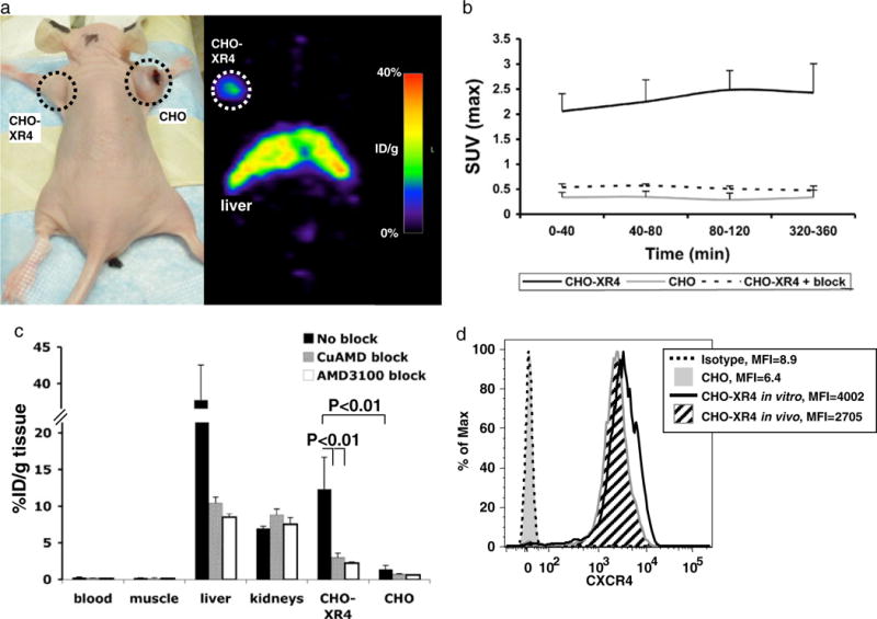
Micro-PET and biodistribution of 64Cu-AMD3100 in CHO-tumor-bearing mice. a Photograph and micro-PET scan of a nude mouse with CXCR4-positive (left) and CXCR4-negative (right) CHO xenografts, injected with 0.925 MBq (25 μCi) of 64Cu-AMD3100 2 weeks after injecting cells and scanned 6 h after injecting the tracer. These results are representative of six mice from two experiments. b Standardized uptake values (SUVmax) were calculated from scans of mice as shown in a, for CXCR4-positive tumors (CHO-XR4, solid line), CXCR4-negative tumors (CHO, gray line), and CXCR4-positive tumors in mice injected with excess unlabeled Cu-AMD3100 (CHO-XR4+ block, dashed line). Each time point is of at least three mice from two experiments. Error bars show positive standard deviations. c Biodistribution of 64Cu-AMD3100 in mice bearing CXCR4-positive (CHO-XR4) and CXCR4-negative (CHO) CHO tumors 2 weeks after injecting the cells and scanned 6 h after injecting the tracer. Data from mice injected without blocking (No block) are in black, with blocking with unlabeled Cu-AMD3100 (CuAMD block) in gray, and with blocking with AMD3100/plerixafor (AMD3100 block) in white. Each group consisted of at least five mice from two experiments. Bracketed horizontal lines indicate significant differences between CHO-XR4 without blockade and the CuAMD3100- and AMD3100-blocked CHO-XR4 and between CHO-XR4 without blockade and CHO without blockade. d Expression of human CXCR4 by cells from CHO-XR4 (angled black lines fill) and CHO (solid gray fill) tumors was evaluated ex vivo by flow cytometry after removing MHC-I H-2Dk-expressing recipient cells from the analysis electronically (see “Materials and Methods”) and compared with the cells grown in vitro (black line) and isotype control staining (dashed line). Mean fluorescent intensities (MFI) are shown. Data are from one CHO-XR4 and one CHO tumor and are representative of results from at least three tumors of each type.