
Figure 1. Vector design.
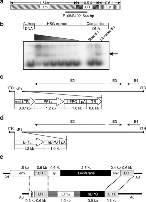
(a) Schematic representation of the 2.7 kilobase (kb) Moloney murine leukemia virus element used in AdLTR-luc,13 and used as a template to amplify 10 different DNA fragments for protein– DNA binding studies with HSG cell nuclear proteins. F105/B102 indicates the position of a 544-base pair (bp) fragment exhibiting the greatest interaction. (b) Autoradiograph of a representative gel shift assay. The interacting DNA–protein complex is denoted by the arrow, which shows that the F105/B102 544-bp fragment has strong and specific binding with increasing concentrations of the HSG cell nuclear protein extract. (c) Schematic depiction of AdLTR2EF1α-hEPO. (d) Schematic depiction of AdEF1α-hEPO. (e) Schematic comparison of the transgene cassettes in AdLTR-luc13 and AdLTR2EF1α-hEPO (reported herein). See text for additional details. Ad, adenovirus; EF1α, elongation factor-1α; env, envelope; hEPO, human erythropoietin; ITR, inverted terminal repeat; LTR, long-terminal repeat; pA, polyadenylation.