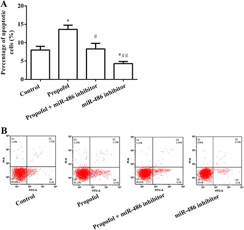
Figure 3. A, Percentages of apoptotic cells in the different groups measured by Annexin V/FITC and PI staining. Apoptotic cells were significantly increased by propofol, but these effects were reversed by transfection with miR-486 inhibitor in H1299 cells. B, Flow cytometry of cell apoptosis. Data are reported as mean ±SD. MiR: microRNA; PI: propidium iodide. *P<0.05 compared to the control group; #P<0.05 compared to the propofol group; # #P<0.01 compared to the propofol group (t-test).

