
Figure 8.
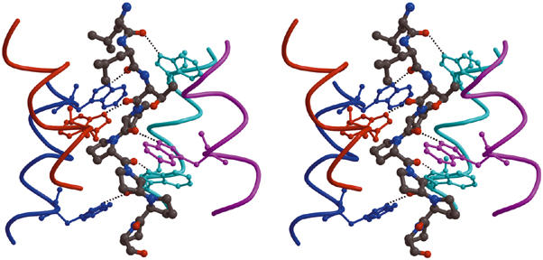
The WAT–PRAD staircase interactions. Stereo view of part of the structure, showing the hydrogen-bond network. The PRAD (gray ball-and-stick format) is surrounded by the WAT helices (depicted as individually color-coded chains). The distal C-terminal Trps (in ball-and-stick format) of each WAT interact with the main-chain carbonyls at the N-terminus of PRAD (top of the figure). For simplicity, only the first six PRAD residues are shown; however, all the main-chain carbonyls of PRAD (1–11) are within H-bonding distance of the Nɛ1 atoms of the Trp indole rings on the WATs.