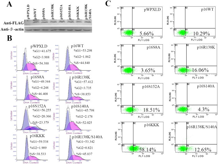
Figure 1. The serine and arginine site mutants of p16 protein exhibited different functions in cell proliferation and apoptosis.
(A) Western blotting analysis of the p16 protein in 293T cells transfected with wild type p16 or mutant p16 expression plasmids, or empty control vector (pWPXLD) as a control. Flow cytometric analysis of cell cycle changes (B) and the apoptosis (C) after transfection of 293T cells with empty control vector (pWPXLD), wild type p16 or mutant p16 expression plasmids. (B) Cells were harvested at 48 h after transfection and stained with PI, analyzed by flow cytometry. (C) Cell apoptosis was measured by flow cytometry after annexin V and propidium iodide (PI) double staining after transfection 48 h.

