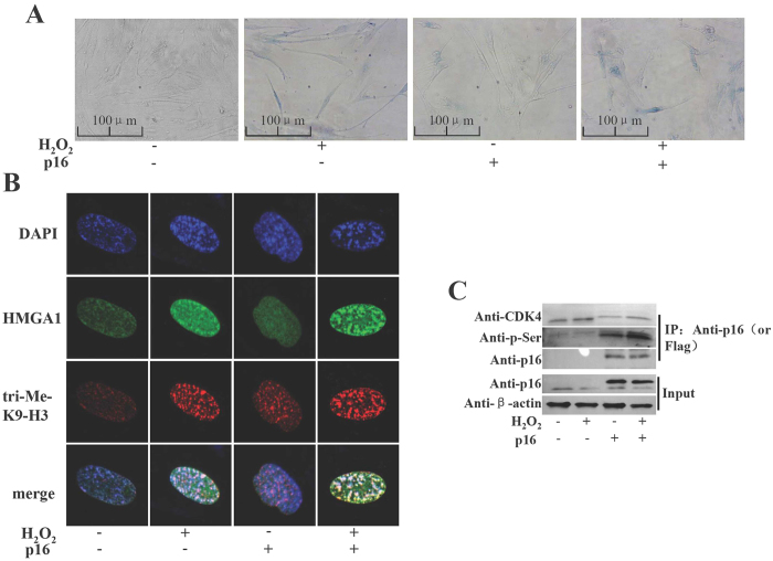
Figure 5. H2O2 induced the senescence and p16 phosphorylation in WI-38 cells.
(A) WI-38 cells were transfected with empty control vector (pWPXLD), or wild type p16 expression plasmids. After 24 h the cells were treated with 1 mM H2O2 for 30 min and then cultured 3 days. The cells were treated with 1 mM H2O2 for 30 min again. After 3 days representative photomicrographs of the SA-β-gal staining are detected under a microscope. (B) Cells were stained with DAPI, and the heterochromatic foci were visualized by fluorescence microscopy. The 3meK9H3 was immunostained in red, and HMGA1 in green. The nuclei were counterstained with DAPI (blue). The cells were visualized under a confocal microscope. (C) the WI-38 cells transfected with plasmids indicated and treated with H2O2 as described above. CoIP with anti-p16 or anti-Flag, and detected with anti-CDK4, anti-p16 or anti-phosphserine antibody. Input: proteins prior to immunoprecipitation.

