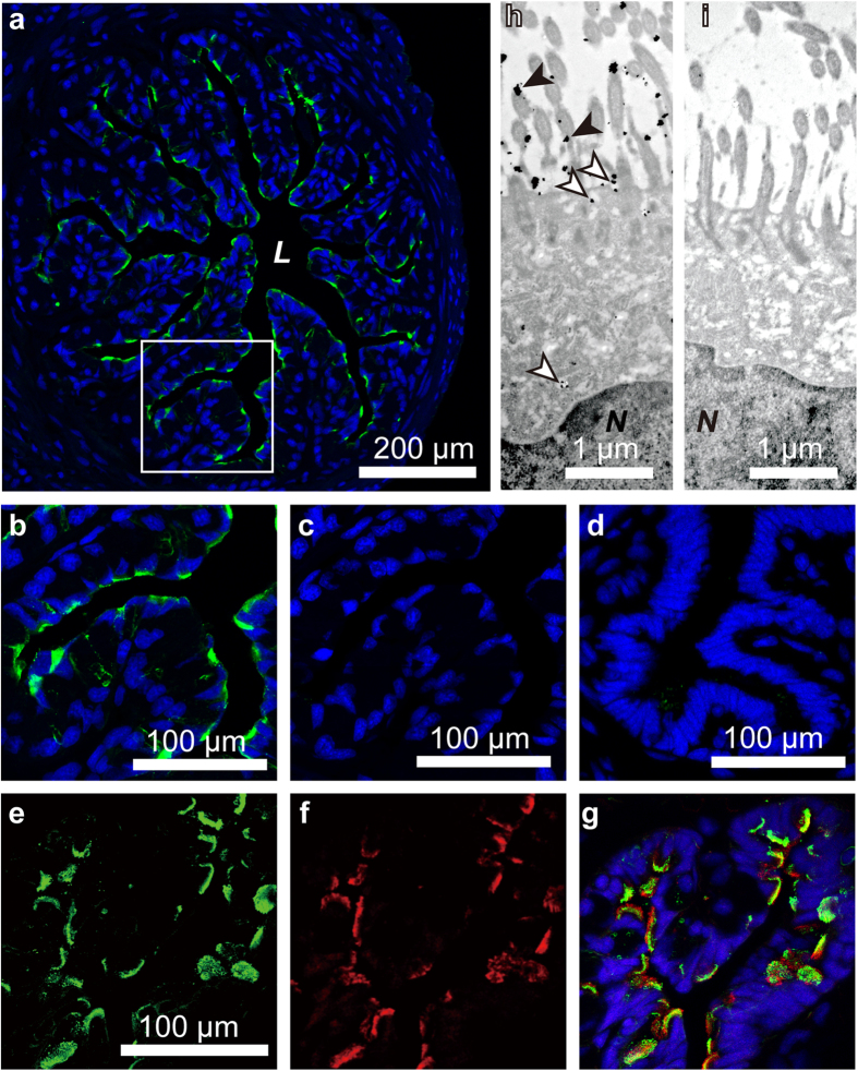
Figure 4. Localization of immunoreactive imorin in the oviduct.
(a) The distribution of immunoreactive imorin (green) in the proximal portion of the oviduct of a sexually developed red-bellied newt (Cynops pyrrhogaster). Nuclei were stained with 4′,-6-diamidino-2-phenylindole (DAPI; blue). (b) Magnified image of the framed area in panel (a). (c) The oviducal section adjacent to the section shown in (b) stained with the anti-imorin antibody preabsorbed with imorin and counterstained with DAPI. (d) A section of the proximal portion of the oviduct of a sexually undeveloped newt that was stained with the imorin antibody and counterstained with DAPI. (e and f) A section of the proximal portion of the oviduct of a sexually developed newt that was double-labelled with anti-imorin antibody (green) and anti-acetylated α-tubulin antibody (red). (g) A merged image of imorin- and acetylated α-tubulin-immunoreactive cells, counterstained with DAPI. (h) A transmission immunoelectron microscopic analysis of the proximal portion of the oviduct of a sexually developed female newt. Imorin-immunoreactive signals were observed mainly in the cytoplasm (outlined arrowheads) of the ciliated cell and on the surface of the cilia (arrowheads). Note that the immunogold particles in the cytoplasm are enveloped in the secretory vesicles. (i) An ultra-thin section of the oviducal portion that is comparable to that shown in (h) stained with anti-imorin antibody preabsorbed with antigen. L, lumen; N, nucleus.

