
Figure 1.
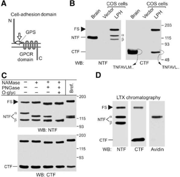
Cellular processing of latrophilin. (A) A typical LNB GPCR. Constitutive cleavage (arrow) creates two fragments corresponding to the cell-adhesion domain and GPCR domain. (B) Analysis of latrophilin expression in COS7 cells. Solubilised receptors from rat brain and COS7 cells transfected with vector or LPH were enriched by α-latrotoxin chromatography and analysed by WB. N-terminal sequences of the native and recombinant CTFs extracted from the gel are shown. (C) Analysis of post-translational modification of latrophilin. Cells were either cultured in the presence of brefeldin A or solubilised and treated with glycosidases, as indicated. For abbreviations of enzymes, see Materials and methods. (D) Identification of surface-exposed species of latrophilin. Live cells were biotinylated, solubilised, enriched on α-latrotoxin column and analysed by WB. Only αNTF is labelled with biotin. Molecular masses are shown on the right (B, C). FS, full-size latrophilin.