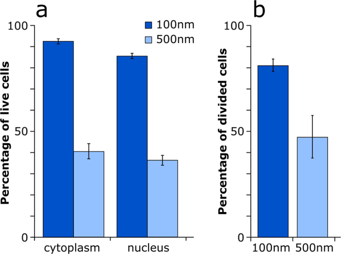
Figure 5. Statistics of cell status 24 hours after the injection of DAF with a 100 nm and 500 nm pipette into the nucleus and cytoplasm.

Prior to injection, the cells were cultivated on μ-Dishes for at least 24 h. For the injection process which took all in all less than an hour, the cell medium was substituted with pre-warmed PBS for a better approach feedback and afterwards again replaced with pre-warmed DMEM and kept in an incubator at 37 °C, 5% CO2. (a) shows viability after the different experiments one day later. For cytoplasmic injection it is 92% (N = 68) for 100 nm and 40% (N = 50) for 500 nm. Injection into the cells nucleus leads to an insignificantly lower viability of 85% (N = 71) and 36% (N = 50) respectively. (b) Comparison of the surviving cells only, with regard to the proliferation percentage. Either we found them alone after 24 hours or we were able to see daughter cells containing the injected dextran in both cells, indicating the proliferation. 81% (N = 116) of the nanoinjected cells divided, whereas only 47% (N = 36) of the cells treated with the 500 nm pipette showed proliferation.
