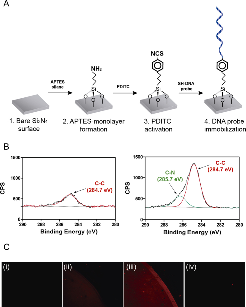
Figure 2. Characterization of the biofunctionalization process on Si3N4 surfaces.
(A) Scheme of the protocol followed for Si3N4 sensor surface biofunctionalization. (B) XPS analysis of the binding energy for C1s bonds in untreated (left) and APTES-modified Si3N4 surfaces (right). (C) Fluorescent signals from the Cy3-Fas57 target for (i) untreated, (ii) PDITC-activated, (iii) complementary DNA-functionalized, and (iv) non-complementary DNA-functionalized sensor surfaces.

