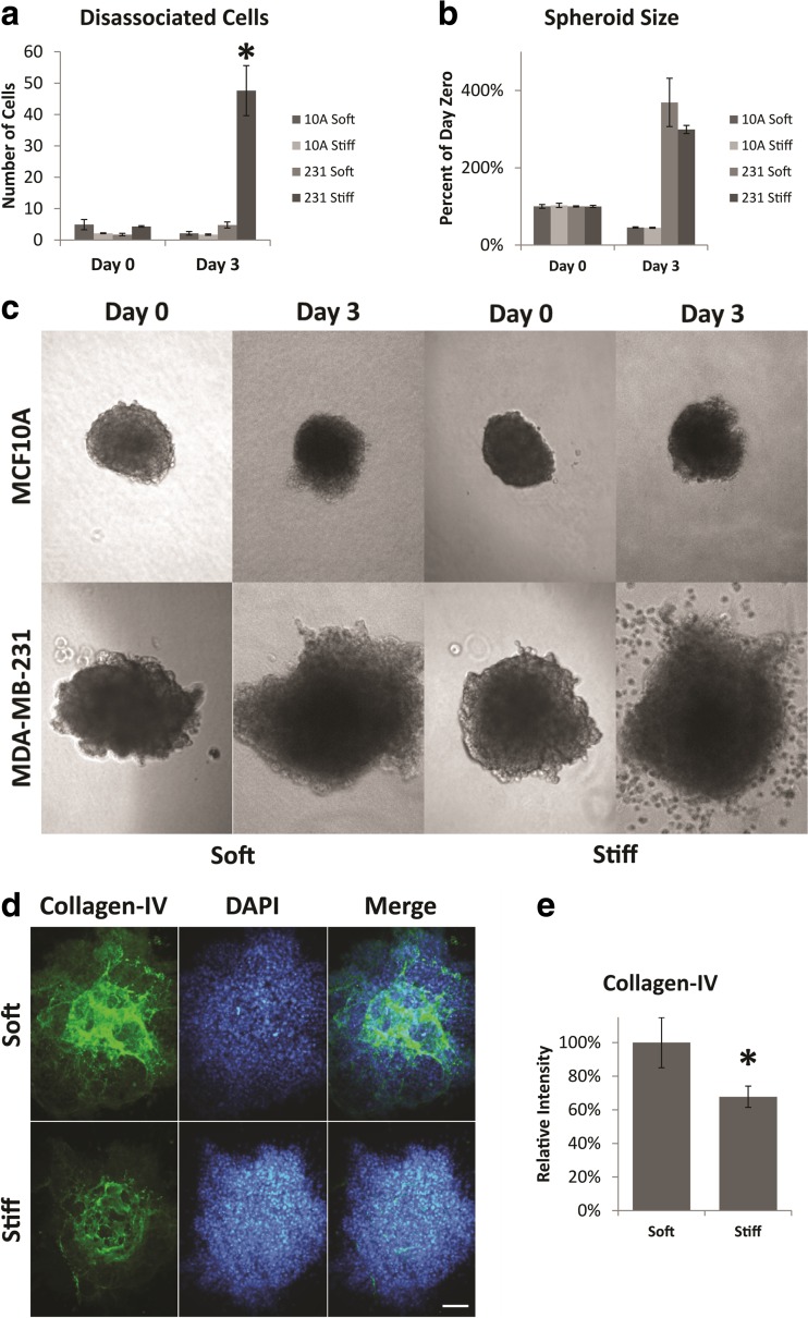
Fig. 2.
Dissemination and size of spheroids. a Number of MCF10A or MDA-MB-231 cells that have dissociated from the main tumor spheroid after 3 days of culture. Disseminated cells were counted as cells which were not in contact with the main spheroid body – including disseminated clusters. b Final size of the tumor spheroid after 3 days of culture. c Representative phase contrast images of spheroids at day 0 and day 3. Images are of the same spheroid at day 0 and day 3. d Day 3 staining of collagen-IV is reduced in stiff hydrogels. e Quantification of collagen-IV staining. Asterisk indicates significant difference from day 0; Student’s t-test p-value <0.05. Error bars show standard error of the mean. All scale bars are 100 μm

