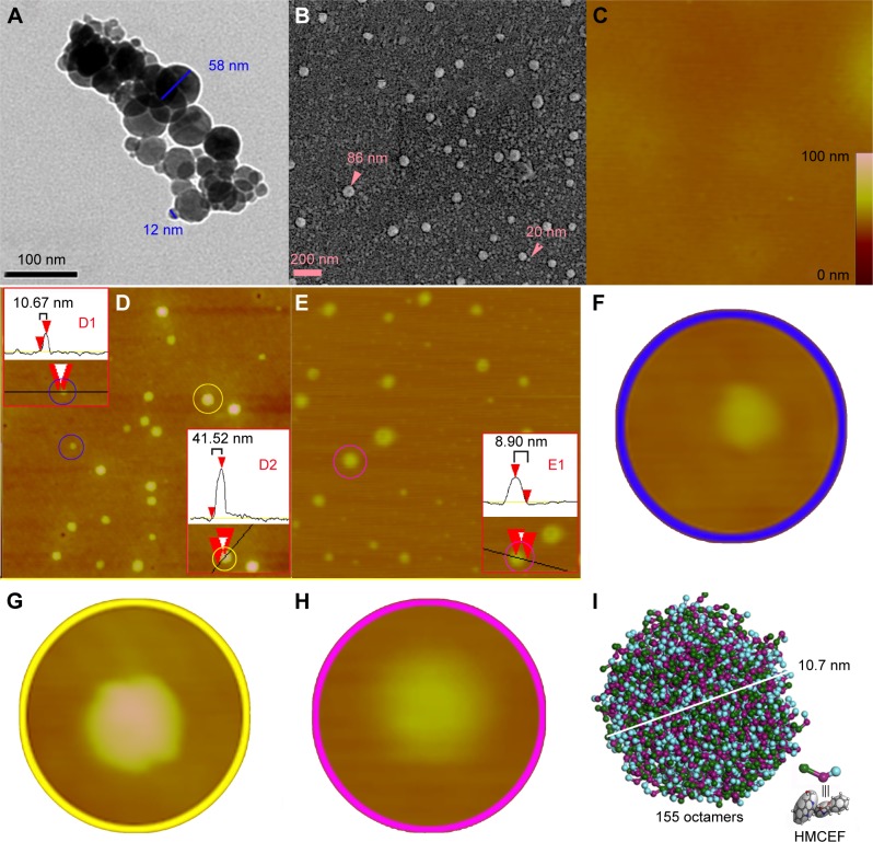
Figure 10.
TEM, SEM and AFM images of HMCEF, as well as the prediction of a nanoparticle of 10.7 nm in diameter by mesoscale simulation.
Notes: (A) TEM image of HMCEF in ultrapure water (pH 7.0, 10−6 M) and amplified nanoparticle of 10 nm in diameter, which has a porous surface. (B) SEM image of the lyophilized powders from 10−6 M solution of HMCEF in ultrapure water (pH 7.0, 10−6 M) and amplified nanoparticle, which has a porous surface. (C) AFM image of rat plasma alone, and gives no comparable particle. (D) AFM image of HMCEF in ultrapure water (10−6 M). D1 scales the particle inside the blue ring; D2 scales the particle inside the yellow ring, and the magnified area identified by the red triangles. Red triangles are plot markers; black lines are grid cursors. (E) AFM image of HMCEF in rat plasma (10−6 M). E1 scales the particle inside the pink ring, and the magnified area identified by the red triangles. In D and E the red triangles are plot markers and the black lines are grid cursors. (F) Magnification of the particle inside the blue ring of D to show its morphology. (G) Magnification of the particle inside the yellow ring of D to show its morphology. (H) Magnification of the particle inside the pink ring of E to show its morphology. (I) Mesoscale simulation predicts that a nanoparticle of HMCEF contains 206 octamers.
Abbreviations: AFM, atomic force microscopy; HMCEF, N-(3-hydroxymethyl-β-carboline-1-yl-ethyl-2-yl)-l-Phe; SEM, scanning electron microscopy; TEM, transmission electron microscopy.

