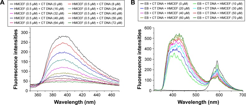
Figure 4.
Fluorescence spectra of HMCEF.
Notes: (A) Fluorescence spectra of HMCEF in PBS buffer (concentration: 0.5 μM, pH =7.4, λex =254 nm) explain the fluorescence quenching induced by 10 μL of CT DNA (final concentrations: 0, 8, 16, 24, 32, 40, 48, 56, 64 and 72 μM) in PBS. (B) Fluorescence spectrum of EB plus CT DNA and HMCEF (the concentration of EB is 5 μM, the concentration of DNA is 10 μM and the concentrations of HMCEF are 0, 10, 20, 30, 40, 50, 60 and 70 μM, respectively).
Abbreviations: CT DNA, calf thymus DNA; EB, ethidium bromide; HMCEF, N-(3-hydroxymethyl-β-carboline-1-yl-ethyl-2-yl)-l-Phe; PBS, phosphate-buffered saline.

