Fig. 4.

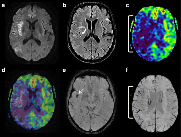
A 76-year-old patient seen in an emergency setting for right brachiofacial motor deficit 3 h after symptom onset. DWI (a), FLAIR (b), ASL (c), ASL/DWI fusion (d), and SWI (e and f) sequences in the axial plane. a Acute right superficial sylvian infarction. b The FLAIR sequence fails to visualise any infarct zone. Slow circulation in the cortical branch of the right middle cerebral artery, hypersignal (curved arrow). c Blue zone (white parentheses) visualises a wide right sylvian zone of hypoperfusion. d Mismatch: DWI hypersignal and ASL hypoperfusion. e Susceptibility vessel sign in the M2 segment of the right middle cerebral artery, thrombus. f Right sylvian (white parentheses) brush sign; the extension is the same as the hypoperfusion zone visible on the ASL sequence (image c)
