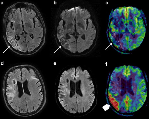
Fig. 8.

Brain MRI in an 85-year-old patient with a history of right sylvian ischaemic stroke presenting with recurrent left hemibody deficiency. FLAIR (a and d), DWI (b and e), and ASL/DWI fusion (c and f) sequences in the axial plane. a, b, c Sequelar right posterior sylvian zone with no sign of recent ischaemia (white arrows). d, e, f Hyperperfusion zone bordering the superior part of the cavity (arrowhead), without FLAIR or DWI anomaly, related to a partial seizure on ischaemic sequelae
